How EM Fields Affect Brain Functions
A research-backed exploration of how electromagnetic stimulation and neuromodulation can alter, disrupt, or modulate specific sensory perceptions and cognitive processes — from vision to taste.
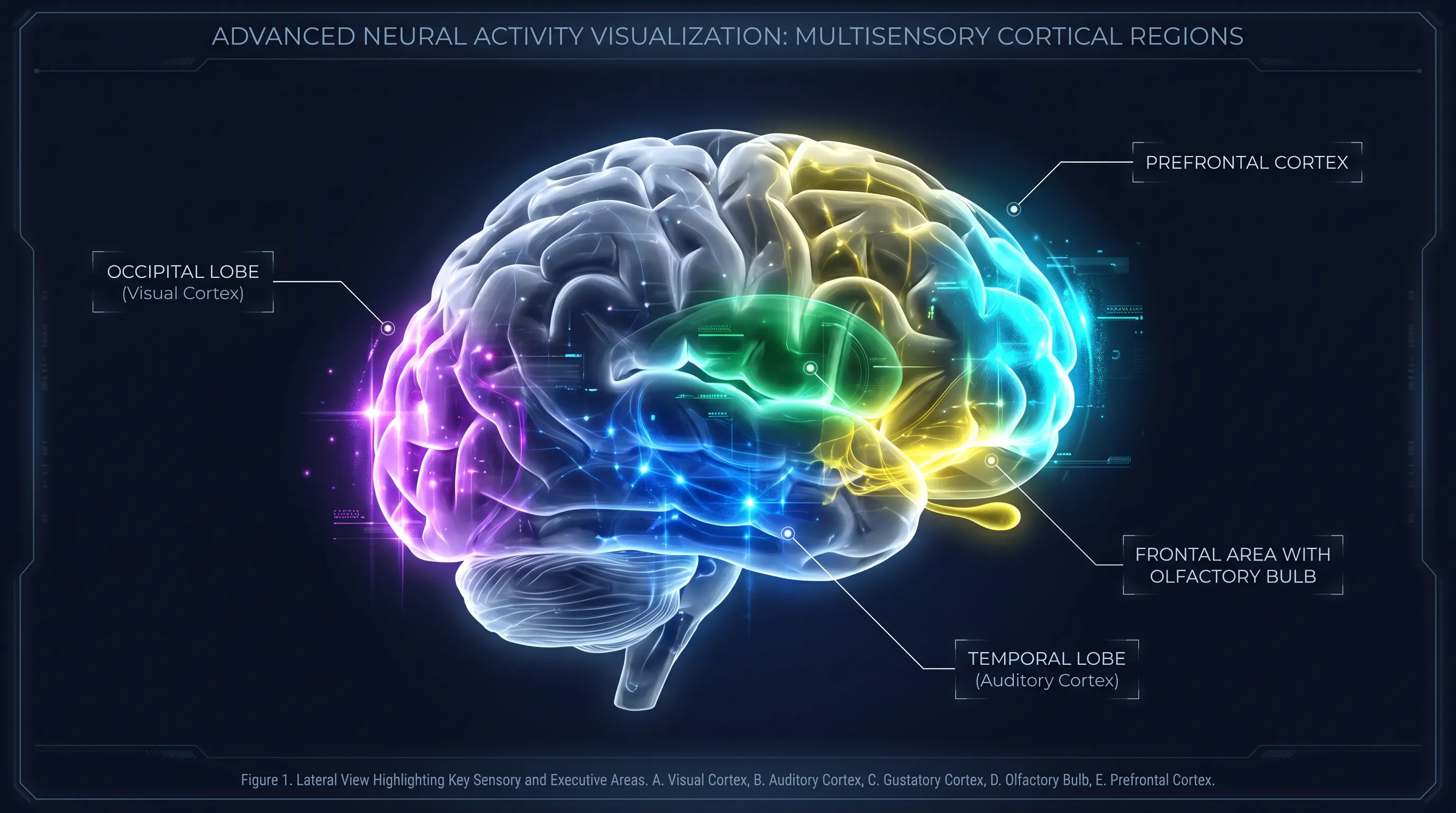
Visual System — Phosphenes & Cortical Disruption
The visual system is one of the most well-studied targets of transcranial magnetic stimulation. TMS applied to the occipital cortex (V1/V2) can induce phosphenes — illusory flashes of light perceived in the absence of visual input — demonstrating direct electromagnetic influence on conscious visual perception.[28]
When TMS is delivered at precise timing windows (~80–120 ms after visual stimulus onset), it can suppress visual awareness entirely, creating transient scotomas (blind spots) in the visual field. This demonstrates that magnetic pulses can both create and destroy visual perception.[29]
Phosphene Induction
Single-pulse TMS over V1/V2 at ~60–80% machine output reliably induces phosphenes. These are perceived as brief flashes of light, typically in the contralateral visual field. The phosphene threshold serves as a standard measure of visual cortex excitability.[28]
Visual Suppression
At supra-threshold intensities, TMS can block the perception of real visual stimuli. When applied 80–100 ms after stimulus presentation, subjects report seeing nothing — a complete interruption of visual processing. This temporal window corresponds to feedback processing from higher visual areas to V1.[29]
Sub-threshold Enhancement
Paradoxically, TMS at intensities just below the phosphene threshold can enhance visual detection. Abrahamyan et al. (2011) showed that subthreshold stimulation at 100–120 ms improved detection of faint stimuli, suggesting TMS-induced neural activity can summate with stimulus-evoked activity.[29]
Key Finding: TMS can both induce visual percepts (phosphenes) and suppress real visual input, with the effect depending on stimulus timing and intensity — demonstrating bidirectional electromagnetic control of vision.
Auditory System — Sound Perception & the Frey Effect
The microwave auditory effect (Frey effect), first documented by Allan Frey in 1962, remains the most dramatic demonstration of electromagnetic influence on auditory perception. Pulsed microwave radiation at frequencies 200 MHz – 3 GHz can induce perceived sounds — clicks, buzzing, or hissing — without any acoustic source.[10]
The mechanism is thermoelastic expansion: rapid, minute heating (~10⁻⁶ °C) of cranial tissue creates pressure waves in the cochlear fluid that are detected by hair cells. The effect requires pulsed, not continuous, radiation and peak power densities of approximately 40 mW/cm².[11]
Mechanism Parameters
Frequency range: 200 MHz – 3 GHz. Pulse width: 1–100 µs. Peak power: ~40 mW/cm². The sound is perceived even by deaf individuals with intact cochleas, confirming the thermoelastic rather than neurological pathway.
TMS-Induced Auditory Effects
Beyond RF, TMS applied over the temporal cortex (auditory areas) can modulate auditory perception. Repetitive TMS (rTMS) at 1 Hz over the temporoparietal junction is used clinically to reduce auditory hallucinations in schizophrenia, demonstrating direct electromagnetic suppression of unwanted sound perception.[30]
Key Finding: Pulsed microwaves can create sound perception via thermoelastic expansion (Frey effect), while TMS over auditory cortex can suppress it — both demonstrating electromagnetic control of auditory experience.

Memory Blocking — Disrupting Consolidation
Memory formation involves a critical process called consolidation, during which newly encoded information is stabilized and transferred from short-term to long-term storage. TMS can disrupt this process by targeting specific cortical regions during the consolidation window.[31]
Research by Censor et al. and others has demonstrated that applying rTMS to the primary motor cortex (M1) immediately after motor learning can completely prevent the retention of newly acquired skills. The memory is encoded but never consolidated — effectively blocked.[31]
Motor Memory Disruption
rTMS at 1 Hz applied to M1 after learning ballistic finger movements completely abolished retention of motor improvements. The disruption was time-dependent: TMS applied within the first 6 hours after learning was effective, but not after overnight consolidation.[31]
Somatosensory Cortex Role
Bhatt et al. (2019) showed that continuous theta-burst stimulation (cTBS) to the somatosensory cortex — not the motor cortex — almost entirely eliminated retention of force-field adaptation learning. This revealed that memory consolidation involves sensory cortices, not just motor areas.[32]
Threat Memory Disruption
Low-frequency rTMS of the dorsolateral prefrontal cortex (dlPFC) can disrupt the consolidation of threat/fear memories. Applied within the early consolidation window, it prevented the persistence and return of conditioned fear responses — a finding with potential implications for PTSD treatment.[33]
Key Finding: Electromagnetic stimulation can selectively block memory consolidation — motor, sensory, and fear memories can all be disrupted if TMS is applied during the critical post-learning consolidation window (0–6 hours).
Cognition Blocking — Disrupting Thought & Decision-Making
The dorsolateral prefrontal cortex (dlPFC)is the brain's executive control center — responsible for working memory, reasoning, planning, and decision-making. TMS disruption of this region produces measurable cognitive impairment.[34]
Knoch et al. (2006) demonstrated that low-frequency rTMS (1 Hz, 15 min) applied to the right dlPFC induced risk-taking behavior in a gambling task. Participants made significantly riskier decisions compared to sham stimulation, showing that electromagnetic disruption of prefrontal function directly alters judgment.[34]
Working Memory Impairment
TMS applied to the left dlPFC disrupts verbal working memory performance. In reading span tests, participants showed decreased word recall under active TMS compared to sham. This demonstrates that the maintenance processing component of working memory depends on intact dlPFC function.[35]
Visual Working Memory
Disrupting the left inferior frontal gyrus (IFG) with TMS impaired visual working memory by reducing the fidelity of neural representations for both stimulus category and goal relevance. This provides causal evidence that prefrontal regions actively maintain and organize information during cognitive tasks.[36]
Decision-Making Disruption
The right dlPFC is critical for suppressing impulsive choices. When disrupted by TMS, subjects consistently chose riskier options — not because they couldn't evaluate risk, but because the inhibitory control mechanism was temporarily disabled.[34]
Key Finding: TMS can disrupt executive function, working memory, and decision-making by targeting the prefrontal cortex — effectively blocking the brain's capacity for rational thought and impulse control.
Olfactory System — Smell Modulation & Disruption
The olfactory system, while less studied than vision or hearing in the context of electromagnetic stimulation, has shown susceptibility to both electrical and magnetic intervention. The olfactory bulb sits at the base of the frontal lobe, making it potentially accessible to transcranial stimulation techniques.
Kumar et al. (2018) demonstrated the feasibility of inducing smell perception through transethmoid electrical stimulation of the olfactory bulbs. In their pilot study, 3 out of 5 subjects reported perceiving a smell during stimulation (1–20 mA at 3.17 Hz), and the perception was reproducible even after medically induced anosmia.[37]
Olfactory Bulb Stimulation
Direct electrical stimulation (1–20 mA, 3.17 Hz) of the olfactory bulbs through the ethmoid sinus reliably induces smell perception. This proof-of-concept opens the pathway toward olfactory implants for anosmia patients.[37]
rTMS for Phantosmia
Henkin et al. (2011) showed that rTMS can treat phantosmia (distorted smell perception) and phantageusia (distorted taste). In 17 patients, 88% responded to rTMS treatment, with 2 patients achieving complete and lasting inhibition of smell distortions for over 5 years.[38]
Cross-Modal Influence
rTMS applied to the visual cortex can improve olfactory discrimination — a surprising finding demonstrating that olfactory perception integrates information from visual processing areas, and disrupting one modality can paradoxically enhance another.[39]
Key Finding: Electromagnetic stimulation can both create and suppress smell perception. Direct olfactory bulb stimulation induces smell, rTMS treats distorted smell, and visual cortex stimulation unexpectedly enhances odor discrimination.
Gustatory System — Taste Perception & Modulation
The gustatory cortex, located primarily in the anterior insula and orbitofrontal cortex, processes taste information and integrates it with smell, texture, and temperature to create flavor perception. Electrical stimulation can directly modulate this system.[40]
Electrical stimulation of the tongue at individual taste thresholds selectively excites gustatory fibers, producing a metallic taste perception. Higher intensity stimulation additionally activates somatosensory areas, showing intensity-dependent activation of human cortical gustatory and somatosensory regions.[40]
Electric Taste Perception
Electrical neuroimaging of event-related potentials (ERPs) reveals that electric taste activates gustatory and somatosensory cortices in an intensity-dependent manner. The perception is consistently described as "metallic" — a direct electromagnetic induction of taste experience.[40]
Subcortical Taste Modulation
Electrical stimulation of the lateral hypothalamus and amygdala can modulate taste neurons in the brainstem (parabrachial nucleus). This bilateral, convergent modulation demonstrates that taste perception can be altered at multiple levels of the neural pathway.[41]
Pharmacological Enhancement
Serotonin reuptake inhibitors (e.g., paroxetine) increase sensitivity to sweet and bitter tastes by modulating the gustatory system's electrochemical signaling. Higher noradrenaline levels reduce the threshold for sour taste perception — demonstrating chemical-electrical interplay in taste modulation.
Key Finding: Taste perception is directly modifiable through electrical stimulation at multiple neural levels — from tongue to cortex. Electric stimulation induces metallic taste, while subcortical stimulation can modulate taste neuron responses.
Implications & Distance Constraints
All documented sensory and cognitive effects require extremely high power densitiesdelivered at close range. TMS coils operate at distances of 1–3 cm from the scalp. Electrical stimulation requires direct contact or implanted electrodes. The Frey effect requires peak power densities of ~40 mW/cm² — orders of magnitude beyond what any satellite can deliver at ground level.
Proximity Requirement
All demonstrated effects require stimulation sources within centimeters of target tissue. No remote, space-based system can replicate these conditions.
Power Gap
The power density gap between satellite signals (~10⁻⁸ W/m²) and biological thresholds (~10⁸ W/m²) spans approximately 10¹⁷ — an insurmountable barrier.
Specificity Challenge
Each sensory modality requires targeting different brain regions with different parameters. Non-invasive precision at distance is physically impossible.
Research Context
These findings demonstrate the brain's electromagnetic sensitivity in controlled lab conditions — they do not support claims of remote sensory manipulation.